Predicting whether chemo will be effective for breast cancer

Breast cancer researchers at UMC Utrecht have discovered that a certain protein can predict whether a course of chemo will be effective for aggressive breast cancer. Women with breast cancer and a high level of FER protein have a greater chance of successful treatment with taxane chemotherapy. The results of this study were published in the renowned scientific journal Cell Reports.

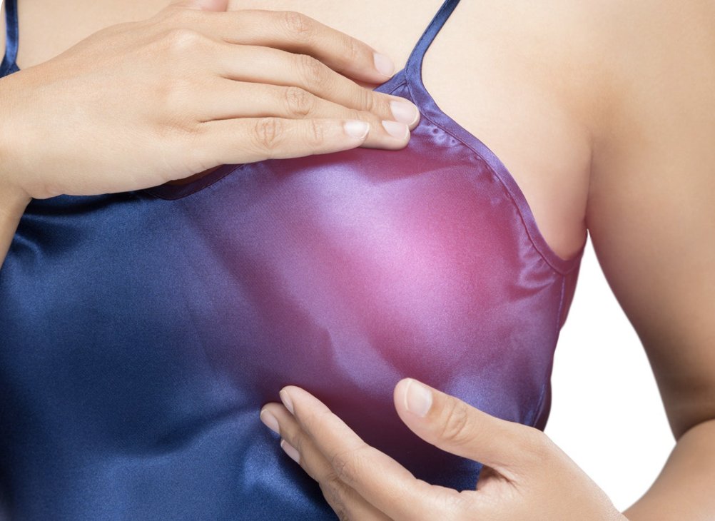
Triple-negative breast cancer is an aggressive type of breast cancer. Women with triple-negative breast cancer often receive treatment with chemotherapy. The goal of the chemotherapy is to reduce the risk of metastasis (spread of the cancer) as far as possible, which increases the chances of survival.

The treatment with chemotherapy is not equally effective for all women. Many women also often suffer unpleasant side effects, such as fatigue, anemia, nausea, and hair loss. “It would be really helpful if we could predict whether a treatment with chemotherapy will be effective,” explains Patrick Derksen, professor of pre-clinical oncology at UMC Utrecht.

High FER protein, greater benefit of chemotherapy

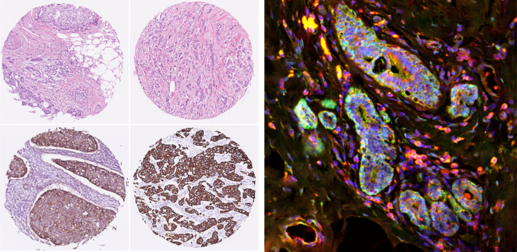
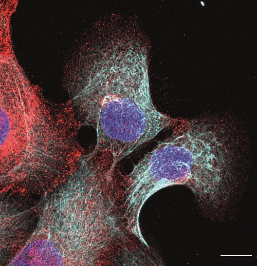
His research group discovered that a certain protein could predict whether a course of chemo will be effective for triple-negative tumors. Women with breast cancer and high levels of the so-called “FER protein” will benefit more from treatment with chemotherapy.

Specifically, they will benefit from chemotherapy with taxanes, a type of medication that slows down cell division. “The FER protein basically recycles other proteins that the cancer cells need to be able to spread,” Derksen explains. “The taxanes slow down this process.”

Test that indicates whether chemotherapy will be beneficial

The researchers are now working on developing a test that demonstrates whether a triple-negative breast tumor has high or low levels of FER protein. High levels of FER equate to a greater probability of the taxane chemotherapy being effective.

Derksen: “We want to start using the test right from the moment of diagnosis, so that we can offer a more tailored treatment. The test is performed in the lab on collected tumor material. We do not need to ask the patients to do anything extra. We will conduct clinical trials on this test, to confirm our prediction and so be able to offer a more personalized and effective treatment.”

Further information

Source: UMCUtrecht

Mechanobiology of Cancer Summer School 2022

Mechanobiology of Cancer Summer School 2022

The MECHANO·CONTROL consortium, led by several research institutions across Europe, is launching the second edition of the “Mechanobiology of Cancer Summer School” which will take place between September 27th – October 1st 2022 at the Eco Resort in La Cerdanya. The aim of the summer school is to provide training on mechanobiology, and specifically its application to cancer. This school will include lectures as well as practical workshops in different techniques and disciplines, ranging from modelling to biomechanics to cancer biology.

The first edition was a great success, both in participation and scientific level, with more than 60 people from 13 different countries.

Mechanobiology of Cancer Summer School 2019

During this second edition, there will be scientific sessions in the morning, mixing 6 keynote speakers with 18 short talks selected from abstract submissions by junior scientists attending the school. In the afternoon, there will be 2-3-hour practical workshops, given by scientists from the MECHANO·CONTROL consortium. The course will also include leisure activities.

Attendance to the Summer School is open to all students, post-docs, and professionals interested, although priority will be given to junior scientists (up to post-doctoral stage).

You can visit the Mechanobiology of Cancer Summer School 2022 website, where you will find more information about the activities that will be held during the summer school, information on how to register, and the deadlines both for the registration and abstract submission.

The 6 confirmed speakers who will attend the summer school are:

  • Dagmar Iber (ETH Zurich)
  • Hans Van Oosterwyck (KU Leuven)
  • Claudia Fischbach (Director of Cornell’s Physical Sciences Oncology)
  • Giorgio Scita (IFOM – The Firc Institute Of Molecular Oncology)
  • Madeleine J. Oudin (Tufts University)
  • Maria Celeste Aragona (University of Copenhagen)

Also, all MECHANO·CONTROL consortium members will be attending the summer school and will be giving some of the whorkshops: Aránzazu del Campo (Leibniz-Institut für Neue Materialien, INM), Sergi Garcia-Manyes (King’s College London, KCL), Pere Roca-Cusachs and Xavier Trepat (Institute for Bioengineering of Catalonia, IBEC), Patrick Derksen, Antoine Khalil and Johan de Rooij (University Medical Center Utrecht, UMCU) and Marino Arroyo (Universitat Politècnica de Catalunya, UPC).

Preliminary list of workshop topics:

  • Vertex modelling mechanics
  • Single molecule mechanics
  • Fundamentals of breast cancer biology
  • Chemistry of tuneable gels

Don’t miss this great networking opportunity, where you will meet and interact with experts in the mechanobiology field and meet new people to collaborate with in the future!


About MECHANO·CONTROL

TheMECHANO·CONTROL project is focused on the mechanical control of biological function.Mechanical forces transmitted through specific molecular bonds drive biological function, and their understanding and control hold an uncharted potential in oncology, regenerative medicine and biomaterial design.

MECHANO·CONTROL proposes to address this challenge by building an interdisciplinary research community with the aim of understanding and controlling cellular mechanics from the molecular to the organism scale. At all stages and scales of the project, it will integrate experimental data with multi-scale computational modelling to establish the rules driving biological response to mechanics and adhesion. With this approach, it aims to explore novel therapeutic approaches beyond the current paradigm in breast cancer treatment. If the partners can understand cancer biomechanics from the single molecule to the whole organ scale, they’ll be able to control mechanical forces to restore healthy cell behaviour and inhibit tumour progression.

Beyond breast cancer, the general principles targeted by this technology will have high applicability in oncology, regenerative medicine, biomaterials and many other biological processes and diseases.

MECHANO·CONTROL is a project funded by the European Commission, within the Future and Emerging Technologies (FET) proactive program.

Project stories: interview with Nimesh Chahare

Project stories: interview with Nimesh Chahare

Multidisciplinarity and collaboration are key to advancing towards new scientific discoveries, and international projects such as Mechano·Control are proof of that. Nimesh Chahare is a Ph.D. student under the supervision of Xavier Trepat and Marino Arroyo, both PIs of the Mechano·Control project.

Nimesh is working on a microfluidic platform to investigate the role of biophysical forces on cells/tissues. The idea came up thanks to a conversation with his colleague Dr. Anabel-Lise Le Roux while discussing new ideas for stretching cells, and they started working right away on this new idea to make it come to life.


To measure the role of biophysical forces, they are developing a microfluidic stretching instrument called MISTI, that can control the biophysical environment via protein patterning, extracellular matrix stiffness, and dynamic stretching. Furthermore, MISTI is compatible with high-resolution microscopy and mechanical measurements.

MISTI could be a critical tool for the mechanobiology community to probe cells and tissues mechanically at various time scales. Why is that? Because it is a flexible system compatible with upright and inverted microscopy, which will make experiments on stretching cells or tissues more accessible for imaging and drug treatments.

“We could also scale this system for high-throughput experiments without live imaging”

Nimesh Chahare, PhD Student at IBEC

One of the goals of the Mechano·Control project is to develop new technologies to manipulate cells and tissues. MISTI would be very useful to unravel the physical principles behind the biological material organization. Trepat and Roca-Cusachs labs’ past work is a perfect illustration of this: from discovering hydraulic fracture in epithelial tissues to identifying critical parameters for mechanobiology such as loading rate for cells [1,2]: all done with stretching of cells and tissues. The enhanced understanding of mechanobiology would provide new insights to solve problems in multiple fields including human physiology and material design.

“These experiments allow us to understand the beauty of nature. They could inspire us to newer ways of thinking. The knowledge we build could help the world design new materials, organize transport systems, or use our setup in ways we couldn’t imagine”

Nimesh Chahare, PhD Student at IBEC

Thanks to the Mechano·Control project Nimesh has met and interacted with people all over the world. In fact, a student from the UK came to Barcelona to work on optimizing the fabrication of MISTI thanks to the project. “This network fosters a collaborative spirit that moves science further” adds Nimesh.


1. Casares, Laura, et al. “Hydraulic fracture during epithelial stretching.” Nature materials 14.3 (2015): 343-351.

2. Andreu, Ion, et al. “The force loading rate drives cell mechanosensing through both reinforcement and cytoskeletal softening.” Nature communications 12.1 (2021): 1-12.

Researcher Xavier Trepat wins the “Constantes y Vitales” award for his pioneering contributions to the mechanobiology field

Researcher Xavier Trepat wins the “Constantes y Vitales” award for his pioneering contributions to the mechanobiology field

Xavier Trepat, ICREA research professor and group leader at the Institute for Bioengineering of Catalonia (IBEC), has been awarded the “Young Talent Award in Biomedical Research”. The ceremony took place at the Palacio de las Alhajas Mamen Mendizábal, Madrid, with the presence of the Minister of Science and Innovation, Diana Morant, and the Chief Executive Officer of Atresmedia, Silvio González. This is the 7th edition of the “Constantes y Vitales” Awards which is aimed at recognizing the 2021 projects in Biomedical Research and Health Prevention.
Xavier Trepat at the awarding ceremony

Xavier Trepat, leader of the research group Integrative cell and tissue dynamics at IBEC, and member of CIBER-BBN, is a recognized expert and pioneer, at an international level, in the field known as mechanobiology, which studies the role of physics in biological systems. Trepat and his team have developed several technologies over the past decade that have made it possible to observe, and measure at the nano-scale, the properties of cells. In doing so, Trepat and his team have contributed to a better understanding of the fundamental mechanisms underlying cell interaction and communication and have reported astonishing discoveries such as that of super elastic cells

But this researcher, an engineer by training, had also a close experience working at a hospital where research was carried out changed the way of understanding science, has also focused on the role of physics in disease. As he stated in a recent article in Nature, the mechanical properties of tissues also play a role in abnormal cell growth, such as in cancer. In Trepat’s own words, “Rigidity makes cancer cells more malignant.” 

And it is that mechanical forces determine biological entities such as cells and organs, both in health and disease and, therefore, as Trepat points out, “Understanding a cell without physics is like trying to write a book with only half the letters of the alphabet”. 

As an example, Trepat and his collaborators recently used mouse stem cells, together with bioengineering and mechanobiology techniques, to measure, for the first time, cellular forces in laboratory mini-intestines. Thanks to this, the team led by Trepat was able to decipher how the inner wall of the intestine folds and moves. The study, published in the journal Nature Cell Biology, opened the doors to understand the bases of diseases such as celiac disease or cancer, and to find solutions for intestinal pathologies through the development of new therapies. 

Awards for an exceptional international career

The European Research Council (ERC) – a European public organization that finances research projects – awarded Xavier Trepat, in 2020, the prestigious ERCAdvancedGrant, endowed with 2.5M euros. Before obtaining this scholarship, the researcher also obtained the “StartingGrant”, “ConsolidatorGrant” and “ProofofConcept” grants, all of them awarded by the ERC, completing the largest series of distinctions at the international level of this entity. 

Previously, in 2015, Trepat was awarded with the Banco Sabadell Award, one of the most important grants in Spain, in the field of health sciences. 

Xavier Trepat has also been one of the few researchers who have managed to publish his work, as first author, in five journals of the Nature family, specifically in NatureNature Physics, Nature Materials, Nature Methods, and Nature Cell Biology

Research uncovers how damaged nucleus re-seals and repairs itself

Research uncovers how damaged nucleus re-seals and repairs itself

The new study shows the molecular mechanisms which re-seal the nucleus’ protective envelope to shield its genetic material.

Within a cell, the nucleus is surrounded by the nuclear envelope – two layers of phospholipid membrane that prevents the nucleus mixing with other parts of the cell, thus protecting the cell’s DNA from outside damage.

However, the nuclear envelope is sometimes broken and ruptured when it undergoes strain. This can occur when the cytoskeleton (a network of fibres acting as a scaffold and give it a stable structure) exerts pressure on the nucleus.

A new study, led by Dr Monica Agromayor of the School of Immunology & Microbial Sciences, illuminates how pressure is relieved from the cytoskeleton.

Uncovering the mechanisms that underpin rupture repair is a fundamental question in cell biology, and a better understanding of this process has broad implications for human diseases such as cancer, cardiovascular disease, or autoimmunity

Dr Monica Agromayor, School of Immunology & Microbial Sciences

Research indicates that a group of proteins known as the ESCRT machinery is responsible for re-sealing the nuclear envelope.

The paper, published in Developmental Cell, focuses on a protein associated with ESCRT named BROX. It shows that BROX is the key protein in relieving pressure from the cytoskeleton, thereby allowing the nuclear envelope to re-seal.

The ESCRT machinery attaches BROX to the site of rupture on the envelope. This allows BROX to bind to the LINC complex – a group of proteins that connect the nuclear envelope to the cytoskeleton, allowing it to apply excessive pressure on the nuclear envelope.

BROX binding leads to LINC being removed from the rupture site. This reduces the stress enforced by the cytoskeleton, allowing the nuclear envelope to repair itself.

The results support growing evidence that the nucleus is an organelle that can interact with its cellular environment, as opposed to just being a protective case for DNA.

Significantly, these results could also have implications for cancer research as a damaged nuclear envelope is associated with cardiovascular diseases and cancer. For example, BROX gene mutations have been found in familial nonmedullary thyroid cancer. Future research can investigate whether BROX mutations contribute to tumours.

Speaking about the team that conducted the research Dr Monica Agromayor said: “This multidisciplinary work shows the importance of collaborative research. Besides the close collaboration with Juan Martin-Serrano, we were fortunate to join forces with the laboratory of Sergi Garcia Manyes from the Physics Department at King’s.”

Read the full paper ‘The ESCRT machinery counteracts Nesprin-2G-mediated mechanical forces during nuclear envelope repair in Developmental Cell.

Original source: KCL

Researchers discover how cellular membranes change curvature depending on BAR proteins

Researchers from both Pere Roca-Cusachs and Marino Arroyo labs, study how BAR proteins, a family of molecules that bind curved cellular membranes, reshape these membranes. Scientists report in the journal Nature Communications, through both experiments and modelling, the dynamics of these membrane reshaping processes that occur both in normal cells or disease scenarios.

Simulations of a membrane tube (600nm diameter) dynamically reshaped by a BAR protein.

The human body is a complex mosaic made up of a very large number of cells with different properties, but they all share a common feature: they possess an external envelope (also called membrane), which is curved at the micrometer and nanometer scales.

During essential cell functions like migration, endo/exocytosis, or when they are deformed by external forces, cell membranes constantly change shape. Here, BAR proteins play a fundamental role. However, the dynamics and mechanochemistry of the process was not well understood until now.

Now, a group of researchers at the Institute for Bioengineering of Catalonia (IBEC) and at the Polytechnical University of Catalonia (UPC) publish new results in the Journal Nature Communications, providing a foundation to understand several processes of membrane deformation, that occur both in normal cell physiology and in disease scenarios. Specifically, researchers describe novel reshaping events of low-curvature membrane structures by BAR proteins, which were previously not considered. Researchers also show how mechanically deforming the membrane triggers a biochemical response mediated by BAR proteins.

The key to understand cell membranes and their curvature 

Experimentally, researchers developed an in vitro system to mechanically deform artificial membranes, expose them to purified BAR proteins, and observed the resulting dynamics by confocal microscopy. In addition, researchers developed theoretical models to understand the process, capturing the dynamics and mechanochemistry of the process. Combining both, experimental and theoretical approaches, researchers also observed that cell membrane deformations depend on initial membrane shape.

Anabel-Lise le Roux (IBEC) and Caterina Tozzi (UPC), co-first authors of the study, explain the importance of having both experimental and theoretical approaches to understand such complex deformation processes.

“The really tight interaction between experiments and modelling was essential to deeply understand a very complex mechanochemical process”.

Anabel-Lise Le Roux and Caterina Tozzi.

The work published in Nature Communications was senior-authored by Marino Arroyo (UPC) and Pere Roca-Cusachs (IBEC/UB).

Reference article: Anabel-Lise Le Roux, Caterina Tozzi, Nikhil Walani , Xarxa Quiroga, Dobryna Zalvidea, Xavier Trepat, Margarita Staykova, Marino Arroyo, Pere Roca-Cusachs. Dynamic Mechanochemical feedback between curved membranes and BAR protein self-organization. Nat Commun 12, 6550 (2021).

Project stories: interview with Thijs Koorman

Project stories: interview with Thijs Koorman

The ultimate goal of the Mechano·Control project is to be able to abrogate breast tumour progression. To do that, it is very important to apply the Mechano·Control findings at different scales, from single molecules and all the way up to the organism and patient level. For that, Thijs Koorman, a senior postdoc at the DerksenLab at the UMC Utrecht, will perform fundamental  and preclinical research within in the Mechano Control project. His main goal is to translate basic findings with therapeutic potential to target invasive breast cancer. With this interview, we are going to take a step forward and get a closer look to the animal and human models, the largest scale of the Mechano·Control consortium.

Dr. Thijs Koorman runs a variety of different tasks. But first, let’s get to know a little bit more about their role in the Mechano·Control consortium. The main aim within the DerksenLab  is to translate the molecular findings within the Mechano·Control project to animal and human models of invasive breast cancer. This group has a fundamental to translational focus. They provide proof of concept in preclinical models of breast cancer and test novel candidate targeted therapeutic interventions identified within Mechano·Control.

With this in mind, Thijs together with Dr. Daan Visser (technician within the Mechano Control consortium) test the therapeutic potential of the identified molecules in the consortium with the hope to obtain pre-clinical data that can be used to ultimately better treat patients suffering from invasive breast cancer.

And how is this translated into the lab?

“We culture mouse and human organotypic cancer models in both 2D and 3D. The models used represent the two types of invasive breast cancer. As such, they harbour all aspects of human invasive breast cancer, which we study using molecular biochemistry and microscopy”, explains Thijs.       

In particular, Thijs studies the molecular and signalling composition of cell adhesion proteins and how disruption of this complex potentiates oncogenic signalling. For this he uses 2D and 3D organotypic tumor models and preclinical mouse models of invasive breast cancer. The group has a deep interest in how loss of mechano-transducing cell-cell and cell-matrix contacts drive to invasive breast cancers, focussing on lobular carcinoma. The groups has generated a breast cancer sample database of over 2000 patients and collected tumour material of over >1000 patients to test molecular markers.

LEFT PANEL: Multiple examples of breast cancer samples, punched from human invasive breast cancers as spotted as a micro-array cores. Top two cores are H&E stainings, bottom two cores are immunohistochemistry (detection of specific proteins in tissues using antibodies). RIGHT PANEL: Triple immunofluorescence-staining image of a developing breast tumor.

Until now, Thijs and the researchers at the Derksen Lab have discovered a molecular mechanism that explains how invasive breast cancer cells may stop dividing and linger in the body for years and what triggers expansion at the metastatic site. Now, they are probing how to target these “sleeping cells” during dissemination and stop the growing cells with therapeutics.

Discovery-based fundamental science is essential to establish the building blocks of therapeutic interventions. “We aim to bridge both aspects, understanding the molecular signalling and discovering or identifying the means to target them. Most importantly, we have the tools to test such in our clinical mouse models and all subtypes of invasive breast cancer. Being able to make these translational steps fast and efficient is a unique aspect of our work” says Thijs.

Thijs also supports the day-to-day supervision of bachelor, master and the Ph.D. students. He also teaches a variety of undergraduate classes at Utrecht University. Thijs also manages the clinical outsourcing and dissemination, including patient advocate involvement. Thijs also holds a leadership position within the COST Action Lobsterpot (CA19138), an initiative of the European Lobular Breast Cancer Consortium (www.elbcc.org).

According to Thijs, being a part of the Mechano·Control consortium is a thrill and a great opportunity to work with colleagues all over the world that work at very different scales. “A single email, text or conversation is sufficient to spark new ideas or endeavours. Combining all of our skills and thinking outside the box is exciting. Not only for your own development, but also to obtain new views on scientific problems. We have a collaborative spirit and were able to test a lot of hypotheses which otherwise would not have been tested.”

Project stories: Interview with Antoine Khalil

In biomedical research, it is important to mimic the conditions that are found in our body. Traditionally, scientists have used 2D in vitro models where they grew cells on flat surfaces. However, as it can obviously be observed, the cells in our body are in three-dimension. So, wouldn’t it be amazing to create 3D models to study diseases, such as breast cancer?

Dr. Antoine Khalil and Mrs. Leonieke den Outer (master student) during confocal microscopy of cancer invasion in 3D matrices

Antoine Khalil is a senior Post-Doc at the UMC Utrecht in the Netherlands, in Johan de Rooij’s group. Their first and very important role at the Mechano·Control consortium was to establish the 3D in vitro models that recapitulate the characteristics of breast cancer in patients. They have used pieces of tumors (organoids) and grew them in 3D extracellular matrix (ECM). The ECM is a complex network of proteins that surround the cells in our body and provide important signals that control cell behavior. Antoine and his colleagues have characterized these cancer organoids and are trying to understand the nature of the extracellular matrix (ECM) that leads to cancer invasion and metastasis.

Working with organoids and 3D ECM makes in vitro studies more reliable, because they retain several aspects of tumor characteristics, and because most of the invasion that takes place in cancer patients, occurs in a 3D environment. The researchers can manipulate these organoids with the aim to stop the ability of cancer cells to become invasive and therefore, this will provide therapeutic opportunities to target the spread of the tumor cells throughout the body.  

“We use a new type of model system for our research that is called organoid culture. Organoids are made from biopsy materials that are isolated from breast tumors in human patients or mouse models. In this way, we can grow a piece of tumor in a controlled environment and study the effects of specific ECM conditions on the induction of tumor cell invasion”, explains Antoine.

Invasion and spread of breast cancer into the surrounding breast tissue often happen in groups of cells (collective invasion). The invasion of the cancer cell groups is driven and guided by few specialized cancer cells called the leader cells. Antoine and his colleagues have identified and fluorescently-tagged molecules that are exclusively expressed in those leader cells. They also found that several of these molecules are activated by changes in the ECM stiffness and tension. Thanks to that, several of these models and markers are being used by the other members and institutions of the Mechano·Control project.

Antoine and his colleagues aim to unravel the molecules that are most important in the transition to an invasive leader cell state and suggest methods to interfere with this process to prevent breast cancer invasion and metastasis.

The “life-time” of a tumor goes through different stages: In the first stage, the tumor grows in an uncontrolled fashion. In this stage the tumor is still confined and the cancer cells remain at their site of origin. At a later stage, some cancer cells within the tumor undergo a transformation that makes them motile and invasive. When this happens, tumor cells can spread to different regions in the body (including lungs, liver and brain) to form secondary tumors (metastases). That is why understanding how invasion is regulated will allow blocking it and this is very important to prevent and reduce metastasis.

It is now very well known that stiffer extracellular matrices surrounding breast tumors strongly associates with breast cancer invasion. “Our major task in the Mechano·control project is to unravel the molecular machinery in breast cancer cells that is essential for ECM sensing and the acquisition of invasive behavior”.

Figure 1. ECM-Dependent invasion of breast cancer organoids. Breast cancer organoids (MMTV-PYMT) embedded in 3D matrigel and collagen I. In both ECM conditions, the basal cells (yellow) are positioned at the tumor-ECM interface. Only in collagen I, the basal cells undergo a transition into protrusive leader cells and drive collective invasion (white arrowheads).

Until now, their data has established that only a pre-defined subset of breast tumor cells (the basal cells) (Figure 1, yellow) can become the leaders of invasion (Figure 1, white arrowheads). These leader cells drive the movement of groups of cancer cells. Antoine’s work shows that becoming a leader cell is triggered by changes in biochemical and mechanical properties of the ECM.

We found that dynamic bidirectional interactions take place between the leader cells and the ECM. Changes in ECM biomechanics induce protrusive behavior in cancer cells, which causes further changes in the ECM that becomes permissive for directional cancer cell movement. We have identified a set of genes whose expression is activated in response to increase in ECM stiffness; many of those genes regulate ECM remodeling and cellular invasive behavior.”

Antoine Khalil, UMCU

This work and much more is what Antoine and his colleagues in de Rooij’s team have achieved within the Mechano·Control project, and we know that there is more exciting and promising research to come until the project ends. For Antoine being a part of the Mechano·Control community was a great opportunity for his work because this project brings together various research fields at very different scales, “Throughout our meetings, conferences and communications we have established strong connections with the partners and learned so much from experts in the cell-ECM interaction field, at the computational, nanoscale and molecular and cellular levels” tells Antoine.

Project Stories: Interview with Samuel Pearson

Project Stories: Interview with Samuel Pearson

Samuel Pearson moved from Australia to Germany, where he works with research groups all over Europe with one common goal: to understand the factors influencing cancer progression. Samuel, Head of Applications in the “Dynamic Biomaterials” division of the Leibniz Institute for New Materials (INM), led by Prof. Aranzazu del Campo, is one of the Mechano·Control members. His work is focused on upscaling the production of polymer materials for 3D cell culture and bringing them towards commercialisation. Let’s take a closer look into his role within the consortium.

To understand how diseases like breast cancer develop, the response of breast cancer cells to different factors need to be studied one by one in isolation in 3D cultures. Samuel Pearson and his team produce synthetic hydrogels for 3D cell encapsulation with the possibility to adjust different parameters. These hydrogels mimic the properties of the natural breast cancer tissue in normal and disease conditions.

A particular interest of his group is the upscaling of the hydrogel precursors using affordable precursors and the design of stable, easy-to-handle compositions. This is the first step in technology transfer from the Mechano·Control project. With Mechano·Control partners, these materials are further developed into predictive breast cancer models.

Another crucial milestone for Samuel and his team is the processability of the hydrogels, which is related to the rate at which they are formed, i.e. the speed of the crosslinking reaction [1] during which cells are retained in a 3D matrix. The group has developed a patented crosslinking approach [2] based on thiol-methyl sulfone reactions which offer very convenient timescales for uniform encapsulation of cells. “In the future, we want to optimize our 3D cell encapsulation systems for automated high throughput methods that will enhance reproducibility and expand the applicability of our platform” explains Samuel. “The figure below shows how basal and luminal cells in breast cancer organoids organise in response to different adhesive ligands in the surrounding polymer network, which relies on consistent network properties” adds Samuel.


Figure 1: Invasive H7 breast cancer organoids were encapsulated in biofunctional hydrogels made from polymers developed at the Leibniz-INM. After 2 days of encapsulation, the effect of different bioactive ligands RGD and DGEA on polarization of the basal cells (stained with K14) and luminal cells (stained with K8) was explored. Scale bar = 50 um. Cell encapsulation and imaging were performed by Dr. Gulistan Kocer.

The materials developed by Samuel and his team play a crucial role in the Mechano·Control consortium. The high level of control and uniformity of the synthetic hydrogels allows to reduce the experimental variability in the biological studies, and this will help to uncover the individual factors that influence cancer progression. Moreover, this technology will not only be of great use for the consortium, but it also offers broad scientific and commercial potential for other 3D tissue culture models.

We are optimising polymer structure and crosslinking chemistry to make hydrogels with very reliable properties. The focus right now is upscaling to allow high throughput studies and moving towards commercialisation

Samuel Pearson

The Mechano·Control project brings together an interdisciplinary research community with the aim to understand and control how cells transmit and detect mechanical forces, from molecules to organs, and all the way up to the organism level. This requires developing and integrating disparate technologies, and each research group involved in the project is an expert in a very specific field and scale: “Each group has its own deep expertise, but because we are working towards a common goal everyone is really motivated to understand the contributions from the other partners, which deepens everyone’s knowledge and experience.”

Adrián de Miguel Jiménez synthesises polymer precursors used for 3D encapsulation of #tumour spheroids, based on the new methyl sulfone-based crosslinking chemistry developed at INM

[1] A crosslink is a bond that links one polymer chain to another, forming a network. Hydrogels are crosslinked networks of hydrophilic polymers that are swollen with water and widely used for 3D cell culture. The crosslink density is a key parameter that determines the mechanical properties of the hydrogel, which can in turn influence the behaviour of encapsulated cells. In the research described above, covalent crosslinks are formed using a patented thiol-methyl sulfone crosslinking reaction.


[2] del Campo, A.; Farrukh, A.; Paez, J. I. Novel Hydrogels. WO2021001203A1, 2021, Priority DE102019117997A 2019-07-03, Filed 2020-06-23, Published 2021-01-07.

Project Stories: Interview with Zanetta Kechagia

The Mechano·Control project aims to understand and control cellular mechanics from molecules to organs, and all the way up to the organism level. With this approach, its interdisciplinary research community will develop specific therapeutic approaches to tackle breast cancer. Zanetta Kechagia joined the Mechano·Control consortium from the first day that the project was launched. She is a postdoc in the team led by Pere Roca-Cusachs at IBEC, who is the principal investigator of the project.

Zanetta is focused on understanding the role of cell mechanics in the cellular and subcellular level. She is particularly interested on how extracellular matrix (ECM) composition and stiffness can jointly influence cell behaviour. “To achieve this, we are using a series of molecular and mechanical perturbations to unravel the role of specific cell-ECM interactions in shaping cellular mechanoresponses” explains Zanetta. 

Mammary gland epithelial cells. In cyan Intermediate filaments and red integrin β4. Scale bar: 20um (Picture by: Zanetta Kechagia)

Currently, she and other researchers at Roca-Cusachs’ lab are trying to identify which cell-ECM interactions can alter the mechanical properties of the cell cytoskeleton. To do so, she uses optical tweezers to perform oscillatory rheological studies, probing cytoskeletal mechanoresponses that are specific for different extracellular molecular components.

“We have found that cells respond to stiffness differently on different ECM substrates and that integrin molecules are key mediators of such responses. By using different ECM coatings, we can probe specific integrin interactions and record changes in the viscoelastic properties of the cytoskeleton over time.” – Zanetta Kechagia, PostDoc at IBEC

Mechanical forces greatly influence cell behaviour, for example in determining whether they differentiate, proliferate, or adopt a malignant phenotype. Understanding how molecular interactions can lead to changes in cell mechanical properties is a fundamental step in order to understand how mechanics control cellular behaviour and eventually tissue responses and cancer progression.

To meet this end, Zanetta is performing these experiments using breast epithelial cell lines with the aim to validate them at the organoid and tissue level, with the help of the other consortium partners. The Mechano·control project covers all scales, starting from single molecule nanotechnology at the smallest scale, to organoids and animal models at the organism scale to uncover cell responses to force at every scale.

Mechano·Control brings together very distinct but complementary research fields and scientists of the highest calibre in their respective fields. For Zanetta, joining the Mechano·Control project has been a great experience so far. “I had the chance to participate in several meetings, visit the partner institutions and discuss my work with our colleagues.  This helped me to acquire new knowledge and new research ideas to rise. One such exceptional experience was the summer school organised by Mechano·Control in 2019, where all the partners had the chance to exchange research ideas and present their work to an international audience.”