This past 2019 has been a great success concerning the outreach activities carried out within the Mechano·Control project. More than 320 people attended talks, workshops, discussions… related to mechanobiology.
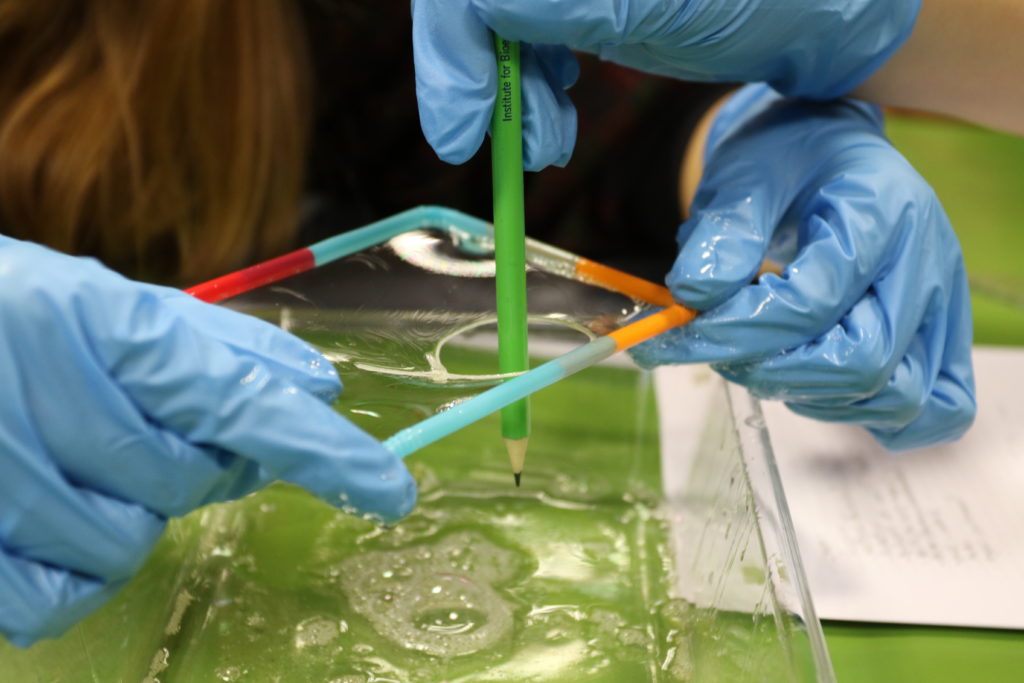
Model on how transmembrane proteins work during a workshop at IBEC
Each year IBEC organises several workshops on mechanobiology where students explore how cells exert forces and they measure them and also create a cell membrane model. This year three schools with 25 students each have participated in this programme. Also, once a year 24 students that participate in a larger programme called “Crazy about bioengineering” come to Pere Roca-Cusachs and Trepat’s lab to do hands-on sessions on how cells perceive the surrounding environment, mechanobiology and biochemical responses.
King’s College
London participated at the 2019 Pint of Science with a talk on how forces are
key to unveiling how life functions with an audience of more than 50 people
from different ages and backgrounds.
UMCU organised
three presentations throughout the year addressed to patients and general
public about their research line on breast cancer, where more than 130 attended
the meetings.
Last but not least, INM also organised two experimental activities at their laboratories reaching 40 students and also mentoring lab practice to 5 secondary school students.
A research team led by the IBEC, in collaboration with the CMR [B], discovers a mechanism that generates binucleated cells.This mechanism has been identified during the regeneration of the heart of the zebrafish, and could be associated with the extraordinary regenerative power of this animal.
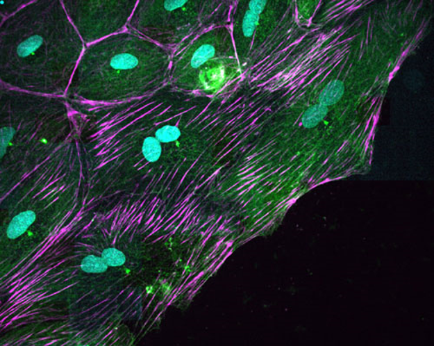
Cells of the epicardium of the zebrafish with two nuclei (in blue)
After an acute heart lesion, such as a myocardial infarction, the human heart is unable to regenerate. The adult cardiac cells cannot grow and divide to replace the damaged ones, and the lesion becomes irreversible. But this does not happen in all animals. A freshwater fish native to Southeast Asia, known as a zebrafish, can completely regenerate its heart even after 20% ventricular amputation.
This extraordinary regenerative capacity has attracted the attention of
researchers from all over the world, who see the range of possibilities that
would be opened up if this mechanism of cell regeneration could be applied in
human therapies.
In an article
published today in the Nature Materials journal, a team of researchers from the
Institute of Bioengineering of Catalonia (IBEC) led by Xavier Trepat, in
collaboration with the Centre for Regenerative Medicine in Barcelona (CMR [B]),
have discovered a surprising mechanism by which zebrafish heart cells move and
divide during regeneration.
Researchers have
focused on the epicardium, which is the layer of cells on the outer surface of
the heart. Although the epicardium cells represent only a small fraction
of the heart’s mass, they play a fundamental role in its regeneration. “The
epicardium is the origin of several of the heart’s cell types, and secretes
biochemical signals that tell the cells what they have to do at all times. It’s
a kind of regeneration ‘hub’”, states Angel Raya, ICREA Researcher and
director of CMRB.
After a heart
lesion, the epicardium cells begin to divide and move en masse to cover the
wound. Researchers have observed that, during this process, the cells become
binucleated: they duplicate the genetic material and separate it into two
nuclei, but they are not divided into two independent cells. “We were very
surprised to discover cells that, instead of having one nucleus, as is the case
in most tissues, they have two nuclei, and each of them contains a copy of the
cell’s DNA” says Trepat, ICREA researcher at IBEC and associate professor of
the University of Barcelona.
Researchers have
discovered that the mechanism by which cells become binucleated has a
biomechanical origin. Once DNA has already separated into two nuclei, most
animal cells form a contractile ring at its centre. As it contracts, this ring
divides the mother cell into two daughter cells. In the case of the heart cells
of the zebrafish, the study shows that the ring adheres to the fibres of its
environment so that it cannot tighten. The result is that the two daughter
cells cannot separate despite having correctly duplicated their DNA.
“Multinucleation
is a well-known phenomenon in cancer, because it is a cause of genetic
instability. In other words, cancer cells lose control of the proteins they
synthesise and behave pathologically. In the case of the heart of zebrafish,
the multinucleation is physiological and does not seem to cause any problem”,
states Marina Uroz, the article’s main author. The next step will be to study
the role of multinucleated cells during the regeneration of the heart and other
organs.
Dr. Trepat and Dr. Raya are part of CIBER-BBN (Biomedical Research Networking Centre in Bioengineering, Biomaterials and Nanomedicine)
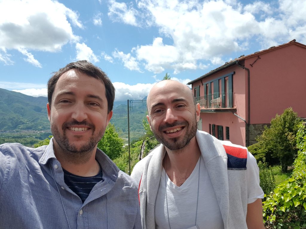
Pere Roca-Cusachs and Antoina Khalil in Lucca during the GRC
Last 5th-10th May was held the 2019 Gordon Research Conference on “Fibronectin, Integrins and Related Molecules” where Pere Roca-Cusachs from IBEC and Antoine Khalil from UMCU presented talks discussing Mechanocontrol results on how mechanical signals and the extracellular matrix regulate cell responses and tumour invasion. During the keynote session titled “Mechanisms and Mechanics of Integrin ECM Connections” Pere Roca-Cusachs gave a talk on “Exploring the Substrate Dependence of Integrin-Mediated Mechanotransduction”. Antoine gave an oral presentation where he described how cell-ECM adhesion regulates the positioning of basal cells and their specification into invasive leader cells during collective invasion of breast cancer organoids.
The Gordon Research Conference was held in Lucca, Italy and is the premier international conference for academic, government and industry scientists interested in understanding how integrins and the extracellular matrix regulate virtually every aspect of cell and tissue function. The program of the conference reflected the interdisciplinary nature of the integrin and extracellular matrix field, spanning different areas of biology from inflammation to mechanobiology, cell migration, stem cells, development, and cancer. During the meeting unpublished data was highlighted and stimulated active discussion among all participants.
The application period opens today until the 8th May 2019, where you can submint an abstract if you are interested in giving a short talk during the summer school.
The application does not guarantee acceptance to the Summer School due to the limited number of participants, an email with the resolution of the applicaton process will be sent on June 15th 2019.
The summer school will be held in La Cerdanya at the Eco-Resort located in Prullans in the Catalan Pyrenees.
The participation fee is 300€ (taxes not included) and includes accomodation in shared double room (from 17th-20th September 2019), full-board, workshops and conferences, leisure activities and shuttle bus from Barcelona to the venue.
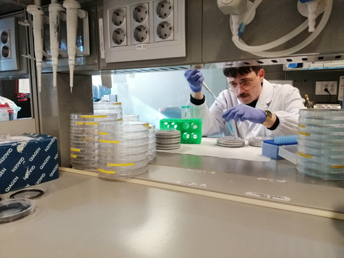
IBEC is
hosting two members from the Mechano·Control network. On the one hand, Dimitri
Kaurin, PhD student from Marino Arroyo group at Universitat Politècnica de
Catalunya (UPC) that will be staying at IBEC for at least one year and on the
other hand, Amy Beedle, postdoc from Sergi Garcia-Manyes at Kings College
London (KCL).
Dimitri Kaurin started his stay at Pere Roca-Cusachs’ laboratory in December 2018 and it is planned to be for at least a year. One of the objectives of Dimitri’s stay is to work on a protocol to study cell-cell adhesion using a controlled system based on lipid bilayers of controlled viscosity. “Using AFM technique, we expect to access some information about cell-cell adhesion under force” says Dimitri. In the context of this research he will also visit Manuel Salmeron laboratory in Glasgow University this march to learn some techniques about functionalizing lipid bilayers with cadherins.
Dimitri Kaurin working in the laboratory at IBEC
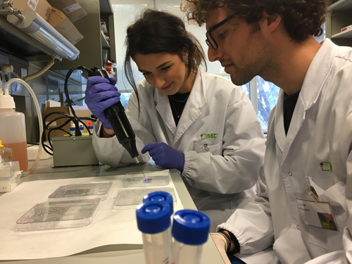
On the other hand, Amy Beedle arrived this past January to Pere Roca-Cusachs’ laboratory. In the Garcia-Manyes lab Amy was looking at how mechanical forces can trigger conformational changes in individual proteins. Here at IBEC, she wants to incorporate the results at the single molecule level with the cellular level, to try to understand how individual bonds and proteins can contribute to cellular mechanosensing. “My aim is to expand my expertise in single molecule force spectroscopy to a larger cellular context” adds Amy.
Amy Beedle working in the laboratory at IBEC
This is the first time that both UPC and KCL teams meet with IBEC to share skills and ideas within the project’s framework.
A review article by Antoine A. Khalil and Johan de Rooij from UMC Utrecht have appeared in the Experimental Cell Research section of Elsevier
Collective invasion drives the spread of multicellular cancer groups, into the normal tissue surrounding several epithelial tumors. Collective invasion recapitulates various aspects of the multicellular organization and collective migration that take place during normal development and repair. Collective migration starts with the specification of leader cells in which a polarized, migratory phenotype is established.
Leader cells initiate and organize the migration of follower cells, to allow the group of cells to move as a cohesive and polarized unit. Leader-follower specification is essential for coordinated and directional collective movement. Forces exerted by cohesive cells represent key signals that dictate multicellular coordination and directionality. Physical forces originate from the contraction of the actomyosin cytoskeleton, which is linked between cells via cadherin-based cell-cell junctions.
The cadherin complex senses and transduces fluctuations in forces into biochemical signals that regulate processes like cell proliferation, motility and polarity. With cadherin junctions being maintained in most collective movements the cadherin complex is ideally positioned to integrate mechanical information into the organization of collective cell migration. Here we discuss the potential roles of cadherin mechanotransduction in the diverse aspects of leader versus follower cell specification during collective migration and neoplastic invasion.
Mechanical forces transmitted through specific molecular bonds drive biological function, and their understanding and control hold an uncharted potential in oncology, regenerative medicine and biomaterial design. However, this potential has not been realised, because it requires developing and integrating disparate technologies to measure and manipulate mechanical and adhesive properties from the nanometre to the metre scale. We propose to address this challenge by building an interdisciplinary research community with the aim of understanding and controlling cellular mechanics from the molecular to the organism scale.
At the nanometric molecular level, we will develop cellular microenvironments enabled by peptidomimetics of cell-cell and cell-matrix ligands, with defined mechanical and adhesive properties that we will dynamically control in time and space trough photo-activation. The properties under force of the molecular bonds involved will be characterized using single-molecule atomic force microscopy and magnetic tweezers.
At the cell-to-organ scale, we will combine controlled microenvironments and interfering strategies with the development of techniques to measure and control mechanical forces and adhesion in cells and tissues, and to evaluate their biological response.
At the organism scale, we will establish how cellular mechanics can be controlled, by targeting specific adhesive interactions, to impair or abrogate breast tumour progression in a mouse model.
At all stages and scales of the project, we will integrate experimental data with multiscale computational modelling to establish the rules driving biological response to mechanics and adhesion. With this approach, we aim to develop specific therapeutic approaches beyond the current paradigm in breast cancer treatment. Beyond breast cancer, the general principles targeted by our technology will have high applicability in oncology, regenerative medicine and biomaterials.