Researchers discover how the intestinal epithelium folds and moves by measuring its forces

Researchers discover how the intestinal epithelium folds and moves by measuring its forces

An international team led by Xavier Trepat at IBEC, with support from “La Caixa Foundation, measures the cellular forces in mini-intestines grown in the laboratory, deciphering how the inner wall of this vital organ folds and moves. The study, published in Nature Cell Biology, opens the doors to a better understanding of the bases of diseases such as celiac disease or cancer, and to the ability to find solutions for gut diseases through the development of new therapies.

Intestine cross section showing its characteristic folded structure. Credit: Amy Engevik

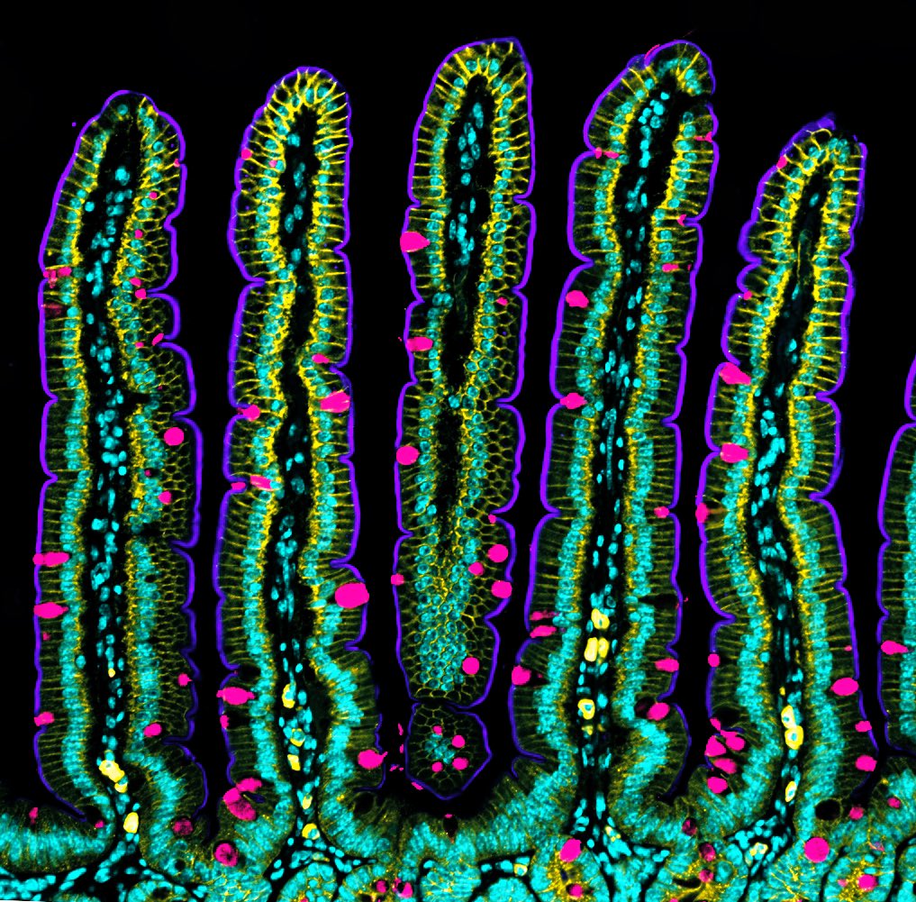
The human intestine is made up of more than 40 square meters of tissue, with a multitude of folds on its internal surface that resemble valleys and mountain peaks in order to increase the absorption of nutrients. The intestine also has the unique characteristic of being in a continuous state of self-renewal. This means that approximately every 5 days all the cells of its inner walls are renewed to guarantee correct intestinal function. Until now, scientists knew that this renewal could take place thanks to stem cells, which are protected in the so-called intestinal crypts, and which give rise to new differentiated cells. However, the process that leads to the concave shape of the crypts and the migration of new cells towards the intestinal peaks was unknown.  

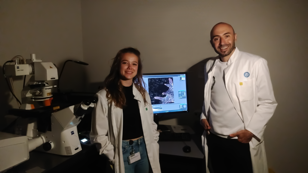
Now, an international team led by Xavier Trepat, ICREA Research Professor and Group Leader at IBEC, in collaboration with the IRB, researchers from the UB and UPC universities in Barcelona, and the Curie Institute of Paris, has deciphered the mechanisms leading the crypts to adopt and maintain their concave shape, and how the migration movement of the cells towards the peaks occurs, without the intestine losing its characteristic folded shape. The study, published in the prestigious journal Nature Cell Biology, has combined computer modelling, led by Marino Arroyo, professor at the UPC, researcher associated with IBEC and member of CIMNE, with experiments with intestinal organoids from mouse cells, and shows that this process is possible thanks to the mechanical forces exerted by the cells. An important part of this study has been supported by the “la Caixa” Foundation within the framework of the CaixaResearch programme. The entity has also awarded a scholarship to the first co-author, Gerardo Ceada, to carry out his PhD at IBEC.

The forces determine and control the shape of the intestine and the movement of the cells

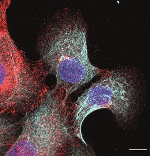
Using mouse stem cells and bioengineering and mechanobiology techniques, researchers have developed mini-intestines, organoids that resemble the three-dimensional structure of peaks and valleys, recapitulating tissue functions in vivo. Using microscopy technologies developed by the same group, researchers carried out high-resolution experiments for the first time that have allowed them to obtain 3D maps showing the forces exerted by each cell. 

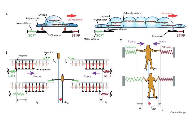
In addition, with this in vitro model, scientists have shown that the movement of new cells to the peak is also controlled by mechanical forces exerted by the cells themselves, specifically by the cytoskeleton, a network of filaments that determines and maintains cell shape. 

Contrary to what was believed up until now, we have been able to determine that it is not the cells of the intestinal crypt that push the new ones up, but that it is the cells at the peak pulling the new ones up, akin to a mountaineer who helps another climber by pulling them up.

Gerardo Ceada (IBEC) 

With this system, we have discovered that the crypt is concave because the cells have more tension on their upper surface than on the bottom, which causes them to adopt a conical shape. When this occurs in several cells next to each other, the result is that the tissue folds, giving rise to a pattern of peaks and valleys.

Carlos Pérez-González, (IBEC and Curie Institute)

The new mini-intestine model will allow further studies of diseases such as cancer, celiac disease or colitis to be conducted in reproducible and real conditions, in which there is an uncontrolled proliferation of stem cells or a destructuring of the folds. In addition, intestinal organoids can be manufactured with human cells and used for the development of new drugs or for the study of the intestinal microbiota.  

Reference article:  C. Pérez-González, G. Ceada, F. Greco, M. Matejčić, M. Gómez-González, N. Castro, A. Menendez, S. Kale, D. Krndija, A. G. Clark, V. Ram Gannavarapu, A. Álvarez-Varela, P. Roca-Cusachs, E. Batlle, D. Matic Vignjevic, M. Arroyo and X. Trepat. Mechanical compartmentalization of the intestinal organoid enables crypt folding and collective cell migration. Nature Cell Biology, 2021.

MechanoControl & Edit: Two new technologies that could change cancer treatment

MechanoControl & Edit: Two new technologies that could change cancer treatment

Two pioneering projects – Mechano-Control and Edit, financed by the European Commission’s FET programme that focus on cancers of the breast and bladder respectively, could provide the necessary tools to better spot and treat them in a non-invasive way.

Read the full article at the FETFX website and get to know more about the projects here

Project stories: Interview with Antoine Khalil

In biomedical research, it is important to mimic the conditions that are found in our body. Traditionally, scientists have used 2D in vitro models where they grew cells on flat surfaces. However, as it can obviously be observed, the cells in our body are in three-dimension. So, wouldn’t it be amazing to create 3D models to study diseases, such as breast cancer?

Dr. Antoine Khalil and Mrs. Leonieke den Outer (master student) during confocal microscopy of cancer invasion in 3D matrices

Antoine Khalil is a senior Post-Doc at the UMC Utrecht in the Netherlands, in Johan de Rooij’s group. Their first and very important role at the Mechano·Control consortium was to establish the 3D in vitro models that recapitulate the characteristics of breast cancer in patients. They have used pieces of tumors (organoids) and grew them in 3D extracellular matrix (ECM). The ECM is a complex network of proteins that surround the cells in our body and provide important signals that control cell behavior. Antoine and his colleagues have characterized these cancer organoids and are trying to understand the nature of the extracellular matrix (ECM) that leads to cancer invasion and metastasis.

Working with organoids and 3D ECM makes in vitro studies more reliable, because they retain several aspects of tumor characteristics, and because most of the invasion that takes place in cancer patients, occurs in a 3D environment. The researchers can manipulate these organoids with the aim to stop the ability of cancer cells to become invasive and therefore, this will provide therapeutic opportunities to target the spread of the tumor cells throughout the body.  

“We use a new type of model system for our research that is called organoid culture. Organoids are made from biopsy materials that are isolated from breast tumors in human patients or mouse models. In this way, we can grow a piece of tumor in a controlled environment and study the effects of specific ECM conditions on the induction of tumor cell invasion”, explains Antoine.

Invasion and spread of breast cancer into the surrounding breast tissue often happen in groups of cells (collective invasion). The invasion of the cancer cell groups is driven and guided by few specialized cancer cells called the leader cells. Antoine and his colleagues have identified and fluorescently-tagged molecules that are exclusively expressed in those leader cells. They also found that several of these molecules are activated by changes in the ECM stiffness and tension. Thanks to that, several of these models and markers are being used by the other members and institutions of the Mechano·Control project.

Antoine and his colleagues aim to unravel the molecules that are most important in the transition to an invasive leader cell state and suggest methods to interfere with this process to prevent breast cancer invasion and metastasis.

The “life-time” of a tumor goes through different stages: In the first stage, the tumor grows in an uncontrolled fashion. In this stage the tumor is still confined and the cancer cells remain at their site of origin. At a later stage, some cancer cells within the tumor undergo a transformation that makes them motile and invasive. When this happens, tumor cells can spread to different regions in the body (including lungs, liver and brain) to form secondary tumors (metastases). That is why understanding how invasion is regulated will allow blocking it and this is very important to prevent and reduce metastasis.

It is now very well known that stiffer extracellular matrices surrounding breast tumors strongly associates with breast cancer invasion. “Our major task in the Mechano·control project is to unravel the molecular machinery in breast cancer cells that is essential for ECM sensing and the acquisition of invasive behavior”.

Figure 1. ECM-Dependent invasion of breast cancer organoids. Breast cancer organoids (MMTV-PYMT) embedded in 3D matrigel and collagen I. In both ECM conditions, the basal cells (yellow) are positioned at the tumor-ECM interface. Only in collagen I, the basal cells undergo a transition into protrusive leader cells and drive collective invasion (white arrowheads).

Until now, their data has established that only a pre-defined subset of breast tumor cells (the basal cells) (Figure 1, yellow) can become the leaders of invasion (Figure 1, white arrowheads). These leader cells drive the movement of groups of cancer cells. Antoine’s work shows that becoming a leader cell is triggered by changes in biochemical and mechanical properties of the ECM.

We found that dynamic bidirectional interactions take place between the leader cells and the ECM. Changes in ECM biomechanics induce protrusive behavior in cancer cells, which causes further changes in the ECM that becomes permissive for directional cancer cell movement. We have identified a set of genes whose expression is activated in response to increase in ECM stiffness; many of those genes regulate ECM remodeling and cellular invasive behavior.”

Antoine Khalil, UMCU

This work and much more is what Antoine and his colleagues in de Rooij’s team have achieved within the Mechano·Control project, and we know that there is more exciting and promising research to come until the project ends. For Antoine being a part of the Mechano·Control community was a great opportunity for his work because this project brings together various research fields at very different scales, “Throughout our meetings, conferences and communications we have established strong connections with the partners and learned so much from experts in the cell-ECM interaction field, at the computational, nanoscale and molecular and cellular levels” tells Antoine.

Project Stories: Interview with Samuel Pearson

Project Stories: Interview with Samuel Pearson

Samuel Pearson moved from Australia to Germany, where he works with research groups all over Europe with one common goal: to understand the factors influencing cancer progression. Samuel, Head of Applications in the “Dynamic Biomaterials” division of the Leibniz Institute for New Materials (INM), led by Prof. Aranzazu del Campo, is one of the Mechano·Control members. His work is focused on upscaling the production of polymer materials for 3D cell culture and bringing them towards commercialisation. Let’s take a closer look into his role within the consortium.

To understand how diseases like breast cancer develop, the response of breast cancer cells to different factors need to be studied one by one in isolation in 3D cultures. Samuel Pearson and his team produce synthetic hydrogels for 3D cell encapsulation with the possibility to adjust different parameters. These hydrogels mimic the properties of the natural breast cancer tissue in normal and disease conditions.

A particular interest of his group is the upscaling of the hydrogel precursors using affordable precursors and the design of stable, easy-to-handle compositions. This is the first step in technology transfer from the Mechano·Control project. With Mechano·Control partners, these materials are further developed into predictive breast cancer models.

Another crucial milestone for Samuel and his team is the processability of the hydrogels, which is related to the rate at which they are formed, i.e. the speed of the crosslinking reaction [1] during which cells are retained in a 3D matrix. The group has developed a patented crosslinking approach [2] based on thiol-methyl sulfone reactions which offer very convenient timescales for uniform encapsulation of cells. “In the future, we want to optimize our 3D cell encapsulation systems for automated high throughput methods that will enhance reproducibility and expand the applicability of our platform” explains Samuel. “The figure below shows how basal and luminal cells in breast cancer organoids organise in response to different adhesive ligands in the surrounding polymer network, which relies on consistent network properties” adds Samuel.


Figure 1: Invasive H7 breast cancer organoids were encapsulated in biofunctional hydrogels made from polymers developed at the Leibniz-INM. After 2 days of encapsulation, the effect of different bioactive ligands RGD and DGEA on polarization of the basal cells (stained with K14) and luminal cells (stained with K8) was explored. Scale bar = 50 um. Cell encapsulation and imaging were performed by Dr. Gulistan Kocer.

The materials developed by Samuel and his team play a crucial role in the Mechano·Control consortium. The high level of control and uniformity of the synthetic hydrogels allows to reduce the experimental variability in the biological studies, and this will help to uncover the individual factors that influence cancer progression. Moreover, this technology will not only be of great use for the consortium, but it also offers broad scientific and commercial potential for other 3D tissue culture models.

We are optimising polymer structure and crosslinking chemistry to make hydrogels with very reliable properties. The focus right now is upscaling to allow high throughput studies and moving towards commercialisation

Samuel Pearson

The Mechano·Control project brings together an interdisciplinary research community with the aim to understand and control how cells transmit and detect mechanical forces, from molecules to organs, and all the way up to the organism level. This requires developing and integrating disparate technologies, and each research group involved in the project is an expert in a very specific field and scale: “Each group has its own deep expertise, but because we are working towards a common goal everyone is really motivated to understand the contributions from the other partners, which deepens everyone’s knowledge and experience.”

Adrián de Miguel Jiménez synthesises polymer precursors used for 3D encapsulation of #tumour spheroids, based on the new methyl sulfone-based crosslinking chemistry developed at INM

[1] A crosslink is a bond that links one polymer chain to another, forming a network. Hydrogels are crosslinked networks of hydrophilic polymers that are swollen with water and widely used for 3D cell culture. The crosslink density is a key parameter that determines the mechanical properties of the hydrogel, which can in turn influence the behaviour of encapsulated cells. In the research described above, covalent crosslinks are formed using a patented thiol-methyl sulfone crosslinking reaction.


[2] del Campo, A.; Farrukh, A.; Paez, J. I. Novel Hydrogels. WO2021001203A1, 2021, Priority DE102019117997A 2019-07-03, Filed 2020-06-23, Published 2021-01-07.

Marino Arroyo, PI of the Mechano·Control project is honoured with an ICREA Academia award

The Catalan Institution for Research and Advanced Studies (ICREA) recognizes the excellent research of Professor Marino Arroyo, who wins for the third time the ICREA Academia award. Over the next five years he will receive a grant in recognition of the research excellence done in his laboratory to continue developing research projects on mechanobiology.

Since 2008, the ICREA Academy program’s goal is to boost and reward the excellence of research at the public universities of Catalonia. It focuses on forefront researchers who are in an active and expansive phase of their career and contributes to intensifying their research. Thirty researchers were recognized in the last call in 2020, including Professor Marino Arroyo.  

Professor Marino Arroyo is an associated researcher at IBEC and member of the Department of Civil and Environmental Engineering and the Mathematics Institute of the Universitat Politècnica de Catalunya (UPC)-BarcelonaTech (IMTech) of the UPC, where he leads the Mechanics of Soft and Living Interfaces laboratory from the Research program on Cell and Tissue Mechanobiology. His research goal is to develop mathematical and computational models to understand the mechanics of biological interfaces, cells and tissues, that is, the small-scale mechanics of materials and biological systems. 

This is the third time Professor Marino Arroyo receives this award, the other two in 2008 and 2015. This new ICREA grant will support research related to the rational manipulation of active living materials and the engineering of new bionic and bioinspired materials, especially materials with extreme reversible deformability. Mechanical aspects of living organisms are a very relevant research field since it plays a crucial role in several biological phenomena, as the development of tumours in cancerous tissues and organs morphogenesis.  

The ICREA Academy programme is funded by the Catalan Government with 5 million per year and grants selected renowned researchers, over a five-year period, with 40.000 EUR per year to boost their research activities. In 2020, the last call of this programme, 30 researchers from different Catalan universities have been awarded, over the 196 applications received. Since the creation of this programme in 2008, 274 researchers already benefited from these grants.  

Project Stories: Interview with Zanetta Kechagia

The Mechano·Control project aims to understand and control cellular mechanics from molecules to organs, and all the way up to the organism level. With this approach, its interdisciplinary research community will develop specific therapeutic approaches to tackle breast cancer. Zanetta Kechagia joined the Mechano·Control consortium from the first day that the project was launched. She is a postdoc in the team led by Pere Roca-Cusachs at IBEC, who is the principal investigator of the project.

Zanetta is focused on understanding the role of cell mechanics in the cellular and subcellular level. She is particularly interested on how extracellular matrix (ECM) composition and stiffness can jointly influence cell behaviour. “To achieve this, we are using a series of molecular and mechanical perturbations to unravel the role of specific cell-ECM interactions in shaping cellular mechanoresponses” explains Zanetta. 

Mammary gland epithelial cells. In cyan Intermediate filaments and red integrin β4. Scale bar: 20um (Picture by: Zanetta Kechagia)

Currently, she and other researchers at Roca-Cusachs’ lab are trying to identify which cell-ECM interactions can alter the mechanical properties of the cell cytoskeleton. To do so, she uses optical tweezers to perform oscillatory rheological studies, probing cytoskeletal mechanoresponses that are specific for different extracellular molecular components.

“We have found that cells respond to stiffness differently on different ECM substrates and that integrin molecules are key mediators of such responses. By using different ECM coatings, we can probe specific integrin interactions and record changes in the viscoelastic properties of the cytoskeleton over time.” – Zanetta Kechagia, PostDoc at IBEC

Mechanical forces greatly influence cell behaviour, for example in determining whether they differentiate, proliferate, or adopt a malignant phenotype. Understanding how molecular interactions can lead to changes in cell mechanical properties is a fundamental step in order to understand how mechanics control cellular behaviour and eventually tissue responses and cancer progression.

To meet this end, Zanetta is performing these experiments using breast epithelial cell lines with the aim to validate them at the organoid and tissue level, with the help of the other consortium partners. The Mechano·control project covers all scales, starting from single molecule nanotechnology at the smallest scale, to organoids and animal models at the organism scale to uncover cell responses to force at every scale.

Mechano·Control brings together very distinct but complementary research fields and scientists of the highest calibre in their respective fields. For Zanetta, joining the Mechano·Control project has been a great experience so far. “I had the chance to participate in several meetings, visit the partner institutions and discuss my work with our colleagues.  This helped me to acquire new knowledge and new research ideas to rise. One such exceptional experience was the summer school organised by Mechano·Control in 2019, where all the partners had the chance to exchange research ideas and present their work to an international audience.”

Mechano·Control featured at the Winter Issue of the EU Research Magazine

Mechano·Control featured at the Winter Issue of the EU Research Magazine

How do forces affect cell behavior? The Mechano·Control project is featured with a one page article in the EU Research Magazine in the “Winter’s Issue: Cellular and Molecular Biology research“.

Pere Roca-Cusachs, PI of the project, explains in this article the aim of the Mechano·Control project which is understanding the mechanical control of the biological function in order to abrogate breast tumour progression.

The consortium addresses this challenge thanks to an interdisciplinary research community with the aim of understanding cancer biomechanics from the single molecule to the whole organ scale. Ultimately, it’s hoped that the project will lead to the development of breast cancer treatments based on the new discoveries.

“Cells change their behaviour when they are in a stiff place; they further stiffen the tissue by secreting more matrix. If you convince the cells that they’re not in a stiff place, then they will stop secreting this, and this may help restore the normal stiffness of the tissue”

Pere Roca-Cusachs

“What happens if we change the composition of the extra-cellular matrix to look more like cancer? What changes?” asks Roca-Cusachs. “We are developing mimics of this matrix, where we can dynamically change their properties. We can make the matrix get stiffer or softer by applying different kinds of light.”

Pere Roca-Cusachs

You can read the article here:

We want to thank the EU Research Magazine for the article (EU Research WIN20/P17.)

Mechano·Control featured at the “Future Tech Week” and “R&I days”

The Mechano·Control project videos have been featured at the “Future Tech week” and at the European Research and Innovation Days event. After submitting the project videos to the “Spotlight Video Contest” of the Future Tech Week, boht videos have been selected to be aired in the session ‘Voices from the Future: EIC Pathfinder projects, stories, people, visions’ held online on the 22th September.

Future Tech Week provides EIC Pathfinder FET projects with a platform from which to blast their exciting findings, results and future paths to innovation to a wide range of stakeholders. It is a platform to showcase all the achievements in fields aligning with the European Commission’s priorities, including Artificial Intelligence and information technology, health and biotech, culture and society, energy and environment, and nanotech and materials. This year’s edition is taking place between 21st – 25th September. Future Tech Week features creative contributions from across Europe and beyond with a focus on Future and Emerging Technologies (FET).

Moreover, on September 22nd and 23rd, Future Tech Week will be part of the flagship European Research and Innovation Days event, within Hub 8 for the European Innovation Council.

The sessions entitled “Voices from the future: Pathfinder stories, people, visions”, organized in the frame of the Future Tech Week, will showcase EIC Pathfinder results from research to their exploitation into the market, through the valuable Keynote speech from Nobel Laureate Professor Edvard I. Moser (GRIDMAP), interview to the EIC Programme Manager Iordanis Arzimanoglou, the Keynote speech by Prof. Jerzy Langer – EIC Pilot Advisory Board member. Several roundtables will discuss about EIC Pathfinder future paths in a wide range of technological trends. The project videos selected by the “Spotlight video contest” will be streamed after Edvard I. Moser Keynote speech.

You can watch the session here

Pere Roca-Cusachs elected member of EMBO

Pere Roca-Cusachs, group leader at the Institute for Bioengineering of Catalonia (IBEC) and associate professor at the Faculty of Medicine of the University of Barcelona (UB), has been chosen to join the European Molecular Biology Organization (EMBO) , a prestigious network that brings together some of the most brilliant researchers in the world. Roca-Cusachs is a pioneer in Europe in the mechanobiology field and in the study of how physical forces affect diseases such as cancer.

Today, the European Molecular Biology Organization (EMBO) has released the names of the researchers who will join the prestigious organization, which has 88 Nobel laureates among its members. In total, 63 life science researchers have been recognized by the EMBO organization for their research career of excellence, among them, Pere Roca-Cusachs, principal investigator of the Institute of Bioengineering of Catalonia (IBEC) and associate professor at the Faculty of Medicine of the University of Barcelona (UB). Pere Roca-Cusachs’ pioneering research at IBEC is focused on the mechanobiology field.

Cutting-edge research in the physics of cancer

As leader of the “Cellular and Molecular Mechanobiology” group at IBEC, Prof. Roca-Cusachs focuses his research to unravel the mechanisms that cells and molecules use to detect and respond to mechanical forces and stimuli, such as, for example, tissue stiffness. These environmental stimuli determine how cells proliferate, differentiate, and move, and regulate processes such as embryonic development, tumour progression, or wound healing.

Since 2017, Pere Roca-Cusachs leads the “Mechano·Control” project, which, with funding from the European Union of more than 7 million euros, seeks to decipher and control how cells transmit and detect mechanical forces. The objective of Mechano·Control is to identify new tools to slow the progression of cancer and especially against breast cancer.

Among other recognitions, Pere Roca-Cusachs received the “Ciutat de Barcelona” award to life sciences 2018, for a study published in the Cell journal, where he identified a mechanism by which tissue rigidity regulates cell survival and proliferation, as well as its implications in diseases such as cancer and liver and lung fibrosis.  In early 2019 he won the award of the “European Biophysical Societies Association (EBSA)” for his contributions in the field of mechanobiology. He is also a member of the “ICREA Academia” program of the Catalan Government.

“Being elected a member of EMBO is a great honour, due to the reputation of the organization and because it is a recognition that comes from my own scientific community. I hope to be worthy of this recognition, not only continuing with our research work, but also contributing to the dissemination and promotion of the importance of research on life sciences that EMBO represents throughout Europe”.

IBEC’s success at EMBO

The EMBO selects new scientists annually, candidate scientists must be nominated by current EMBO members and approved by another five members from different countries and then, they have to receive approval from the entire organization. In this edition, 63 candidacies from 25 different countries have been accepted, 52 of them have become part of the organization and 11 as associate members. Only three Spanish researchers are joining the organizaation and Pere Roca-Cusachs, who back in 2016 was already accepted in the “EMBO Young Investigator Programme”. The EMBO members are excellent scientists who carry out cutting-edge research in the different disciplines of the life sciences, among which there are 88 members who have received Nobel prizes. This new appointment is in addition to Xavier Trepat, also group leader at IBEC, who has been a member of EMBO since 2018.

About EMBO:

EMBO is an organization of more than 1800 leading researchers that promotes excellence in the field of life sciences, both in Europe and worldwide. The organization’s primary goals are to support talented researchers at all stages of their careers, stimulate the exchange of scientific information, and help build a research environment where scientists can achieve their best work. Along with Pere Roca-Cusachs, researchers Maria Dolores Martin-Bermudo from the Pablo de Olavide University and Guillermina López-Bendito from the Institute of Neurosciences of San Juan de Alicante have also been chosen by the organization.

Primer on “Durotaxis”

Molecular clutch model proposed to explain durotaxis.
Molecular clutch model proposed to explain durotaxis (Current Biology)

Xavier Trepat, group leader at IBEC and PI of the Mechano·Control consortium together with Raimon Sunyer, Senior researcher in Trepat’s lab, have written a Primer in Current Biology magazine on “Durotaxis”, a cell migration mechanism that might have a role in several disease states that include the stiffening of tissues.

Embryo development, tumour progression or the immune response against pathogens requires cell migration. Cells are not static, they move and are able to direct their migration, normally guided by spatial gradients in a physicochemical property of the cell microenvironment, such as chemical concentration for example, but it is also guided by the stiffness of their extra-cellular matrix (ECM).

Durotaxis was first reported in 2000 and is the tendency of single cells to follow stiffness gradients. Since it was first described, several studies have been carried out, mostly in vitro, as in vivo remains poorly studied. As technological advances bring new tools to probe ECM stiffness in living tissues, new roles for durotaxis in vivo are likely to emerge.

Durotaxis is normally positive, towards stiff regions, but it has also been observed as negative, from stiff to soft, some examples of this phenomenon are explained in this review.

In this piece, written by Xavier Trepat and Raimon Sunyer, they give an overview on the methods used to study durotaxis both in vitro and in vivo, and on the state of art of the mechanisms of durotaxis, which remains incompletely understood.

The researchers also explain that some cell types do not display significant durotaxis when migrating in isolation, but they durotax efficiently as cohesive clusters. Multicellular clusters can behave as a giant supracell, increasing its sensitivity to mechanical gradients. However, collective durotaxis has not yet been demonstrated in vivo but it is believed that this phenomenon could guide collective migration in pathological processes involving local changes in tissue stiffness. For instance, solid tumours are widely known to be stiffer than the surrounding tissue, which may favour or prevent collective invasion.

In conclusion, in this review the researchers state that durotaxis is emerging as a robust mechanism to drive directed migration of single cells and clusters. Key components include cell-ECM adhesion through molecular clutches and long-range force transmission across the cytoskeleton and cell-cell junctions. It is expected that durotaxis might be the cause of many migratory movements in vivo that are currently unexplained.

Read the full article here: Raimon Sunyer; Xavier Trepat, Durotaxis. Current Biol. 2020, 30, R383-R387